 繁體中文
繁體中文") English (UK)
English (UK) PI

Jyh-Horng Chen, Distinguished Professor, Department of Electrical Engineering, National Taiwan University
Co-PI
Hong-Chang Yang, Professor; Yeun-Chung Chang, Professor; Ang Yun, Clinical Associate Professor; Chia-Hsien Cheng, Associate Professor
In these five years, the aims of MRI core lab are
Develop Wideband sequence with fast, or high spatial resolution MR imaging for early detection of diseases
Design on quantitative susceptibility MR molecular imaging mapping for longitudinal studies of animals/patients
Develop DCE MRI for assessment of lung cancer mice model in variable of drugs and treatment protocol
Work on Translational Medicine on newly developed novel molecular imaging protocols for early detection of cancers
Current Studies
1. Wideband(WB) MRI
Wideband MRI is a technique that utilized wide bandwidth in order to increase the information obtained per unit time. It has the ability to speedup various applications especially those that require large coverage. In other cases, it could be also used for higher spatial resolution or temporal resolution for possible detection of diseases. The versatility of Wideband MRI allows its acceleration ability to be applied to several MRI sequences by means of echo planar imaging, perfusion, image flow, angiography, temperature,T1 imaging, T2 imaging, diffusion and the like.
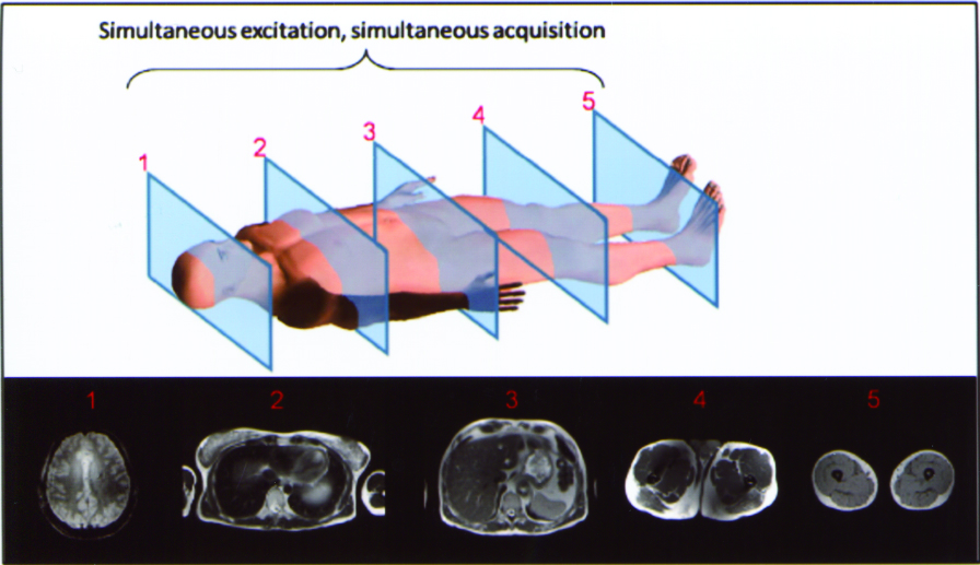
Figure 1. Conceptual illustration of a Wideband MRI human whole body scan with Wideband factor W=5
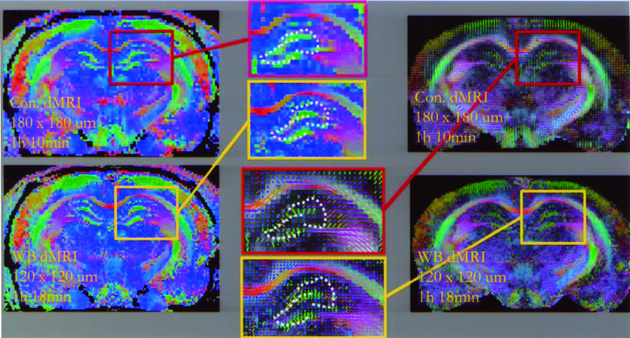
Figure 2. The upper raw: the rat brain diffusion image in resolution of 180 x 180 um, and the bottom raw in high resolution 120 x 120 um using Wideband diffusion technique in the same scanning time
2. Quantitative susceptibility mapping (QSM)
QSM aims to quantify the susceptibility within tissues. Our study proposed a novel methodology for noninvasively detecting cerebral small vessels including veins and venules, called QSM-mMRA. The QSM-mMRA can simultaneously provide the information on cerebral anatomy, micro-vascular architecture, and SvO2, which can be used to evaluate the physiological and functional characteristics of microvascular changes for longitudinal monitoring and therapeutic evaluating with disease model.
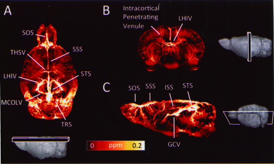
Figure 3. Quantitative visualisation of QSM of a normal rat brain in three orthogonal views. Veins in cortical and internal brain are indicated
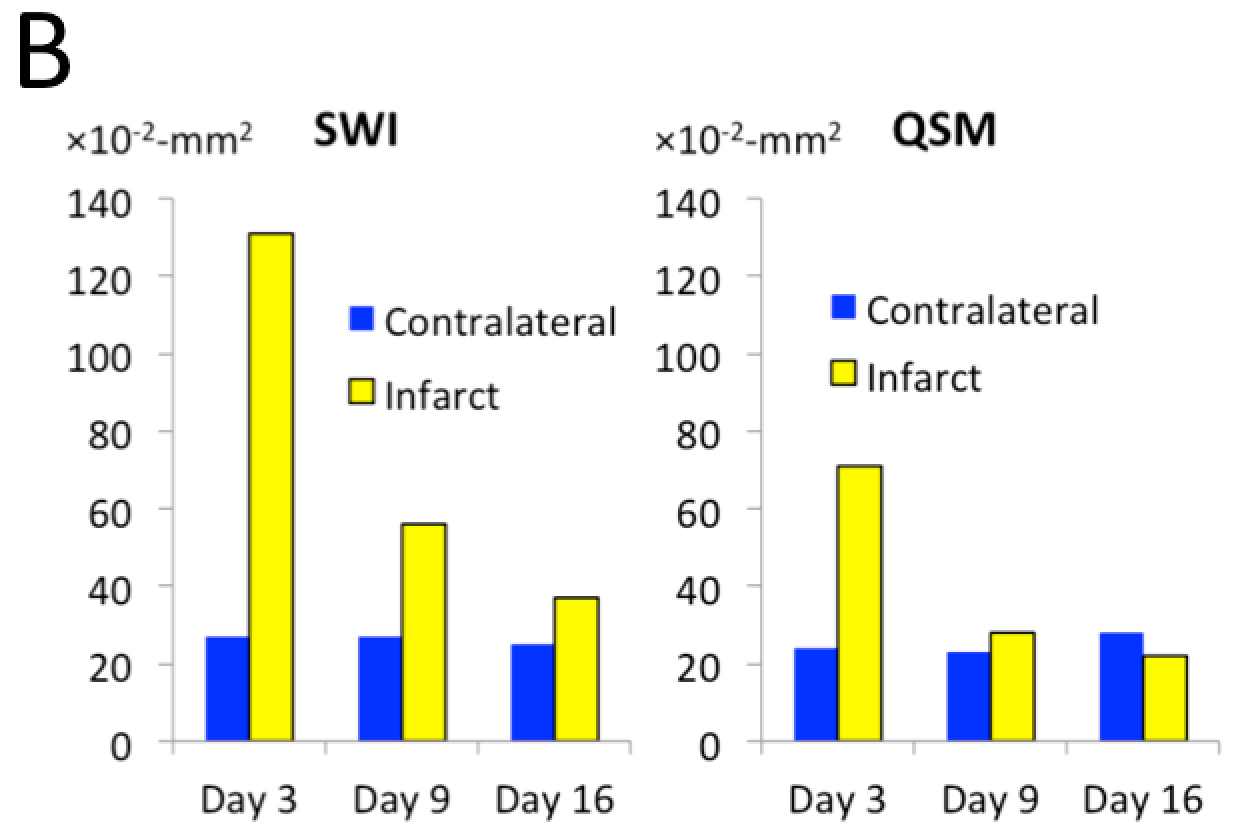
Figure 4. Detection of the rehabilitation of a cortical venule in the rat brain after stroke
Facility
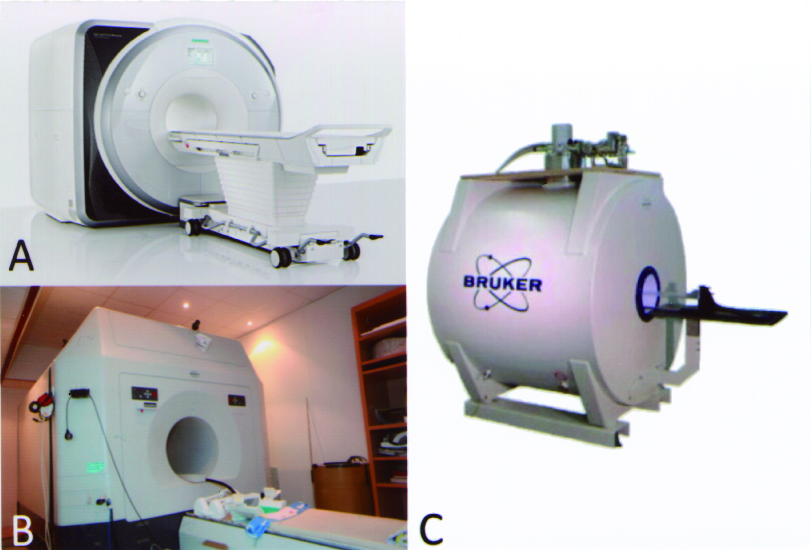
A. Siemens Prisma 3T Human MRI
B. Bruker 3T Human MRI
C. Bruker Biospec 7T Animal MRI