 繁體中文
繁體中文") English (UK)
English (UK) PI

Ruo-Fang Yan, Director, Department of Nuclear Medicine, National Taiwan University Hospital
Co-PI
Ling-Wei Hsin, Associate Professor; Chun-Jung Lin, Associate Professor; Lih-Chu Chiou, Professor; Wei-Chung Hsu, Assistant Professor; Tong-Rong Jan, Professor; Chien-Hung Chen, Associate Professor; Wen-Ming Hsu, Associate Professor; Chih-Hung Hsu, Assistant Professor; Ming-Jang Chiu, Associate Professor; Wuh-Liang Hwu, Professor; Chyng-Yann Shiue, Professor
The goal of the PET core is to use various PET probes in the study of molecular characteristics of biology, physiology, pathology and disease status of the biological systems from small animals to human beings by PET imaging. This non-invasive molecular imaging technique is useful in diagnosis, staging, monitoring and follow up of diseases related to malignancies, degenerative neuronal diseases and ischemic cardiovascular disorders.
Our Core is equipped with two PET/CT scanners for human studies, one PET/CT scanner for small animal research and one cyclotron with full capability of radiopharmaceutical synthesis. Since 2005, the production of PET probes in our lab beyond 18FDG outnumbered almost all other PET Centers in Taiwan.
Hepatocellular carcinoma(HCC) and Alzheimer's Dementia(AD) are now the two main diseases of interest in our lab. We are now performing a human clinical trial in applying 11C-PIB in AD and a multicenter clinical trial in applying 18F-choline in HCC. Furthermore, we started year 2013 using multiple PET probes, 18FDGal and 18F-choline, for the diagnosis of patients with HCC.
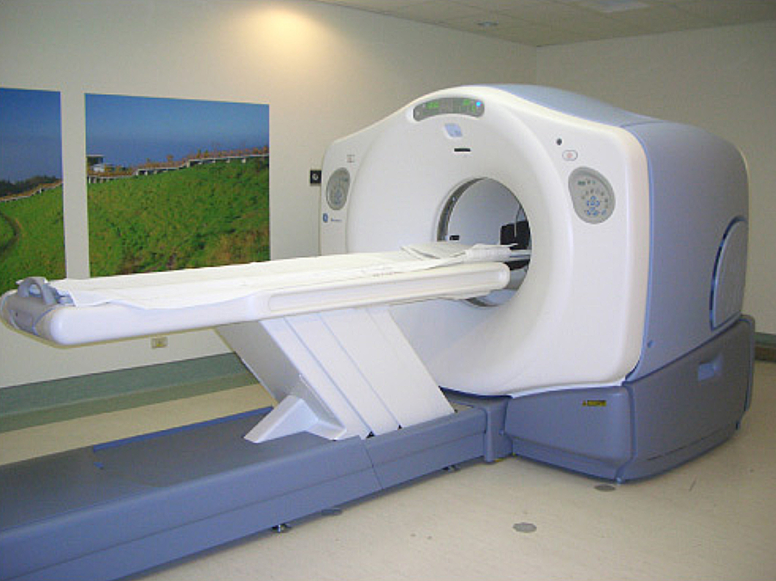
Figure 1. PET/CT scanner
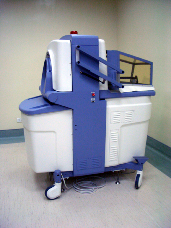
Figure 2. Small animal PET/CT scanner
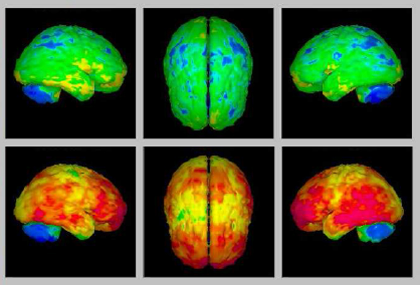
Figure 3. 3D 11C-PiB distribution in healthy control(above) and AD(below)
We use two PET probes(18FDOPA AND 18F-fallypride) and one SPECT probe(99mTc-TRODAT-1) to characterize children with congenital AADC deficiency preparing for gene therapy. The images showed low AADC enzyme content, intact dopamine transporter and intact dopamine D2 receptor. The post therapy image demonstrated increased AADC enzyme effect.
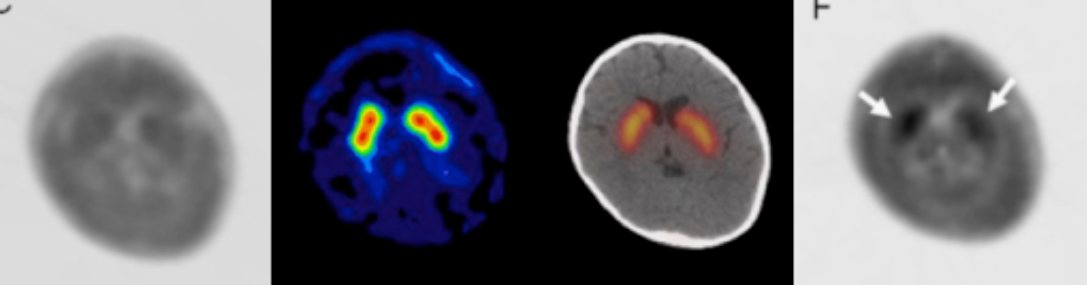
Figure 4. 18F-DOPA before Tx 99mTc-TRODAT 18F-Fallypride 18F-DOPA post Tx
In the era of molecular imaging, we can use several nuclear, both SPECT and PET, probes to target different molecules to define the biochemical phenotypes of diseases. This is true in diseases, such as neuroblastoma, hepatocellular carcinoma, Parkinson's disease, and Alzheimer's dementia. The multiple faces of a disease can be clearly defined using this imaging technique.