 繁體中文
繁體中文") English (UK)
English (UK) PI
Chi-Kuang Sun, Distinguished Professor, Electrical Engineering, Photonics and Optoelectronics, Biomedical Electronics and Bioinformatics, National Taiwan University
Fellow IEEE, OSA, SPIE
Personal Homepage: http://ufo.ee.ntu.edu.tw/
Co-PI
Din-Ping Tsai, Distinguished Professor; Chen-Yuan Dong, Distinguished Professor; Hsuan-Shu Lee, M.D., Professor; Chen-Tung Yen, Professor; Shi-Wei Chu, Professor; Kung-Bin Sung, Associate Professor; Tzu-Ming Liu, Associate Professor; Yi-Hua Liao, M.D., Ph.D; Wei-Hsuan Yu, Assistant Professor; Wen-Jeng Lee, M.D.; Yuan Luo, Assistant Professor; Hwan-Ching Tai, Assistant Professor; Tzung-Dau Wang, Attending Physician (National Taiwan University Hospital)
THE GOALS of the optical imaging core are to improve early diagnosis and risk assessment of diseases, as well as to aid research in preventive and regenerative medicine. We aim to develop innovative optical molecular imaging technologies and establish preclinical and clinical study platforms based on these technologies by combining expertise in fundamental science, engineering and clinical research, with emphases on important health issues including cancer as well as neural, metabolic, and cardiovascular diseases.
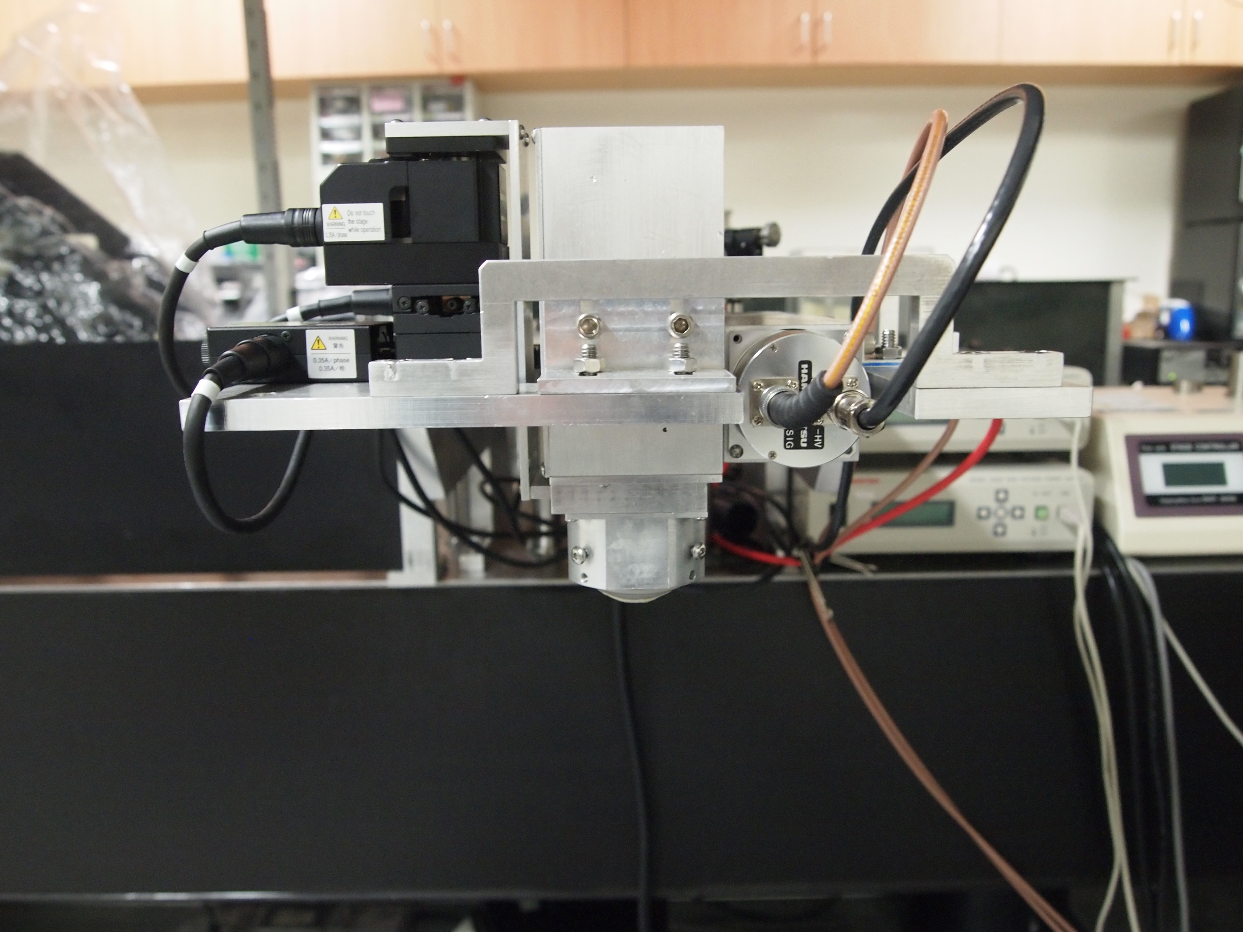
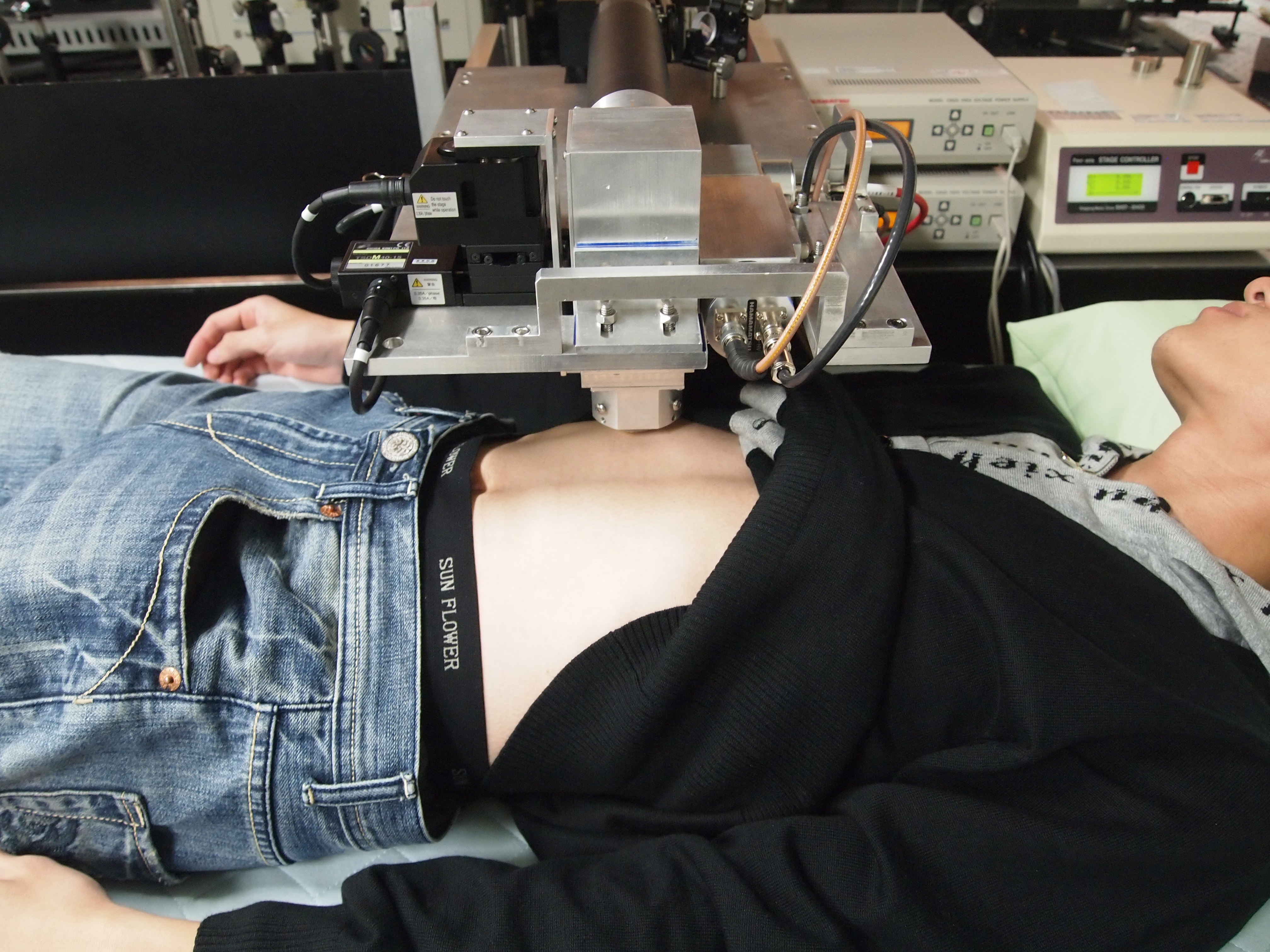
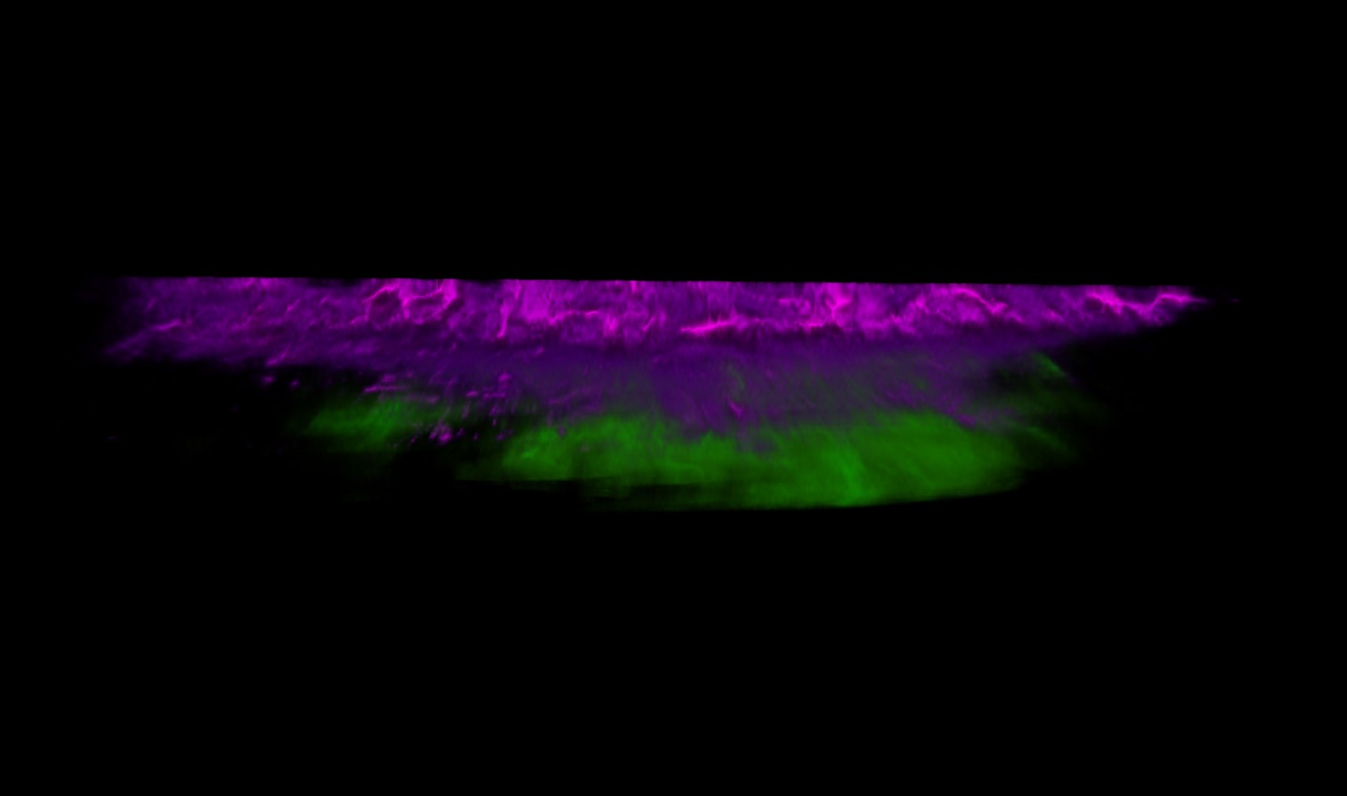
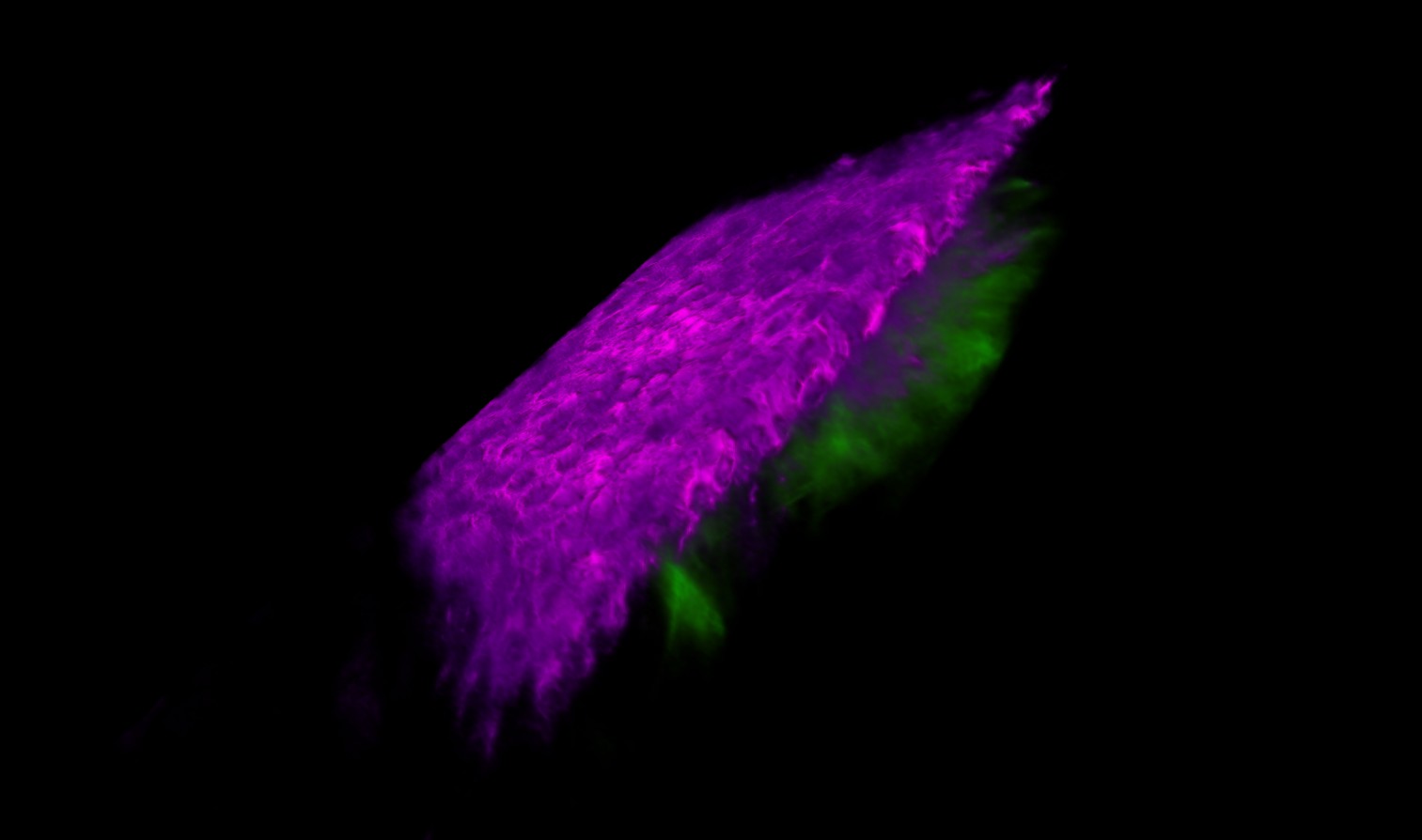
In vivo Harmonic Generation Microscope
|
|
|
|
|
|
|
Movable Hyperspectral Microscope System
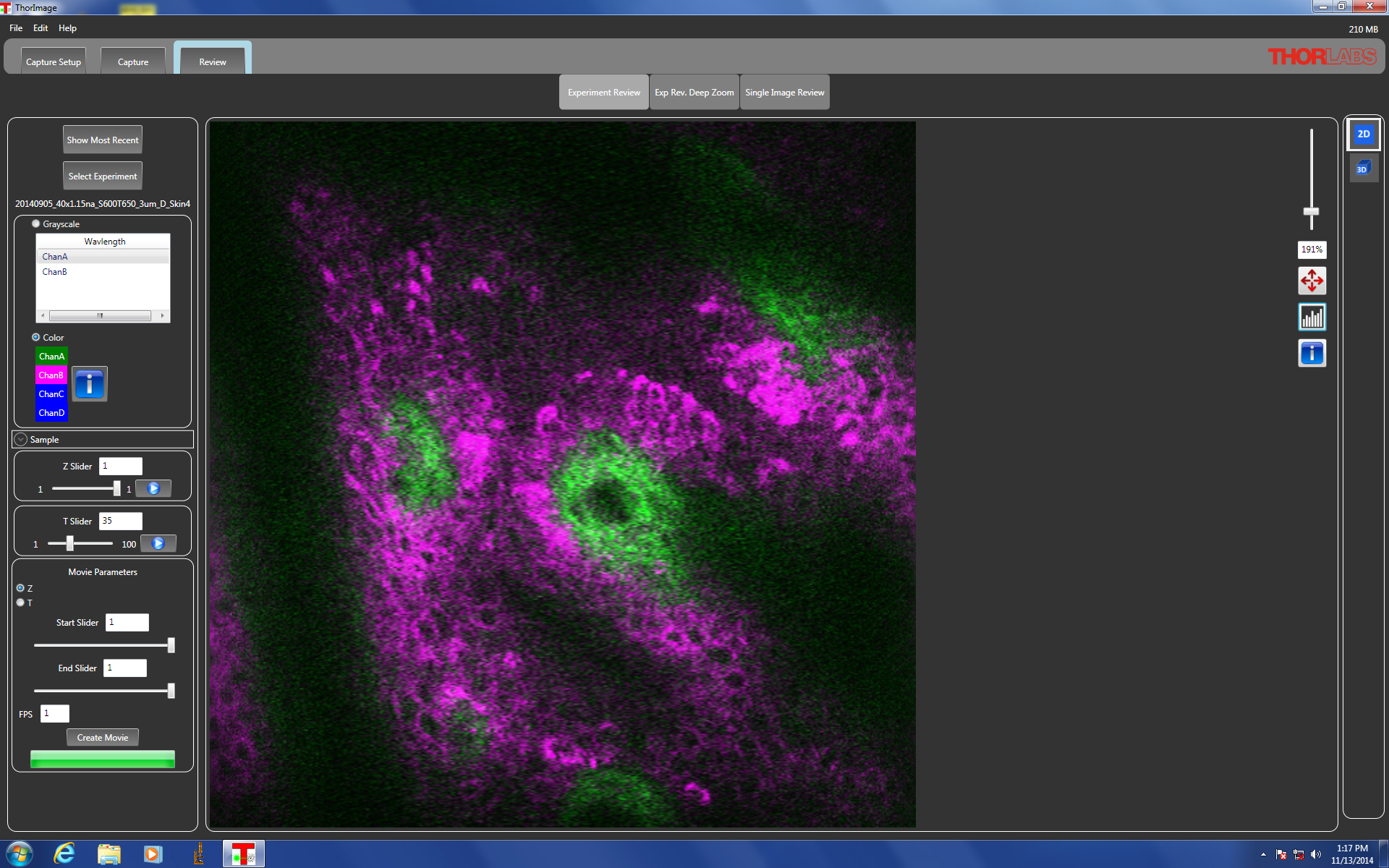
We constructed a movable hyperspectral microscope system for acquiring spatially-resolved reflectance and fluorescence spectra. This system is used to measure oral and esophageal mucosa in vivo to quantify the scattering, absorption and fluorescence properties of the epithelium and underlying lamina propria. We evaluate the performances of the proposed method to distinguish precancerous tissue from normal tissue based on the parameters extracted from in vivo spectra.
LEICA TCS SP5 STED